Copyright
©The Author(s) 2015.
World J Gastroenterol. Dec 14, 2015; 21(46): 13195-13200
Published online Dec 14, 2015. doi: 10.3748/wjg.v21.i46.13195
Published online Dec 14, 2015. doi: 10.3748/wjg.v21.i46.13195
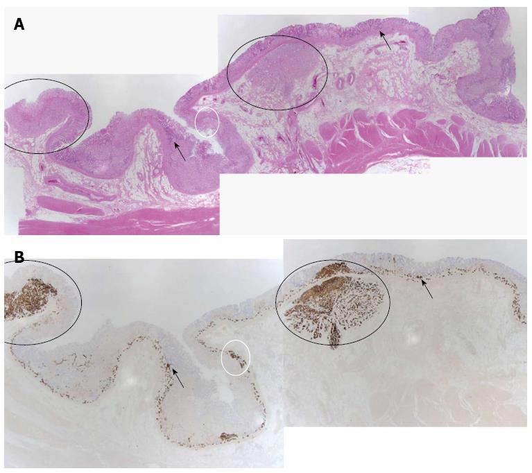
Figure 3 Pathological features.
A: Hematoxylin-eosin staining; B: Immunohistochemical stain (chromogranin stain). The black arrows indicate the lesions of hyperplasia of the neuroendocrine cells. The white circles indicate the lesions of dysplasia of the neuroendocrine cells. The black circles show the lesions of the neuroendocrine tumors.
- Citation: Jung M, Kim JW, Jang JY, Chang YW, Park SH, Kim YH, Kim YW. Recurrent gastric neuroendocrine tumors treated with total gastrectomy. World J Gastroenterol 2015; 21(46): 13195-13200
- URL: https://www.wjgnet.com/1007-9327/full/v21/i46/13195.htm
- DOI: https://dx.doi.org/10.3748/wjg.v21.i46.13195


