Copyright
©The Author(s) 2015.
World J Gastroenterol. Dec 14, 2015; 21(46): 13055-13063
Published online Dec 14, 2015. doi: 10.3748/wjg.v21.i46.13055
Published online Dec 14, 2015. doi: 10.3748/wjg.v21.i46.13055
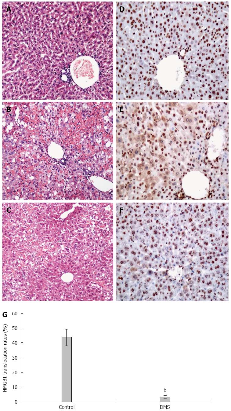
Figure 3 Immune cell infiltration and tissue damage and HMGB1 cytoplasmic translocation in hepatocytes of mice 36 h after D-galactosamine/lipopolysaccharide challenge.
Immune cell infiltration, tissue damage, and HMGB1 cytoplasmic translocation in hepatocytes at 6 and 36 h after the onset of acute liver failure were detected by hematoxylin and eosin staining (A-C) and immunohistochemistry (D-F); magnification × 10 or × 20. A and B: Normal mice; C and D: Acute liver failure mice; E and F: DMS-treated mice; G: Percentage of hepatocytes with HMGB1 cytoplasmic translocation. χ2 = 12.81, bP < 0.01, (3.57% ± 0.83%) vs controls (43.72% ± 5.51%). The mean ± SE of three independent experiments is shown (error bar indicates standard error).
- Citation: Lei YC, Yang LL, Li W, Luo P, Zheng PF. Inhibition of sphingosine kinase 1 ameliorates acute liver failure by reducing high-mobility group box 1 cytoplasmic translocation in liver cells. World J Gastroenterol 2015; 21(46): 13055-13063
- URL: https://www.wjgnet.com/1007-9327/full/v21/i46/13055.htm
- DOI: https://dx.doi.org/10.3748/wjg.v21.i46.13055


