Copyright
©The Author(s) 2015.
World J Gastroenterol. Dec 7, 2015; 21(45): 12778-12786
Published online Dec 7, 2015. doi: 10.3748/wjg.v21.i45.12778
Published online Dec 7, 2015. doi: 10.3748/wjg.v21.i45.12778
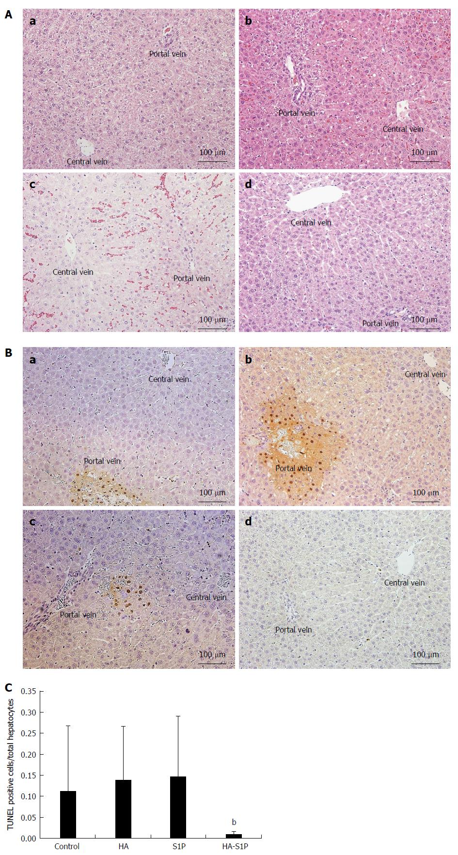
Figure 3 Histological findings.
A: HE staining In the control (a), HA (b), and S1P group (c) histologic examination revealed vacuolation of the hepatocytes and loss of palisade arrangement after 120 min of reperfusion. These findings were not observed in the HA-S1P group (d). (hematoxylin-eosin stain, magnification × 200); B: TUNEL (TdT-mediated dUTP-biotin nick end labeling) assay of the liver tissue and (C) the ratio of TUNEL-positive/total hepatocytes In the control (a), HA (b), and S1P groups (c), TUNEL-positive cells were observed in zone 1 after 120 min of reperfusion. These findings were not observed in the HA-S1P group (d). bP < 0.01 vs the control, HA, and S1P groups. HA: Hyaluronic acid; S1P: Sphingosine 1-phosphate.
- Citation: Sano N, Tamura T, Toriyabe N, Nowatari T, Nakayama K, Tanoi T, Murata S, Sakurai Y, Hyodo M, Fukunaga K, Harashima H, Ohkohchi N. New drug delivery system for liver sinusoidal endothelial cells for ischemia-reperfusion injury. World J Gastroenterol 2015; 21(45): 12778-12786
- URL: https://www.wjgnet.com/1007-9327/full/v21/i45/12778.htm
- DOI: https://dx.doi.org/10.3748/wjg.v21.i45.12778


