Copyright
©The Author(s) 2015.
World J Gastroenterol. Nov 14, 2015; 21(42): 11974-11983
Published online Nov 14, 2015. doi: 10.3748/wjg.v21.i42.11974
Published online Nov 14, 2015. doi: 10.3748/wjg.v21.i42.11974
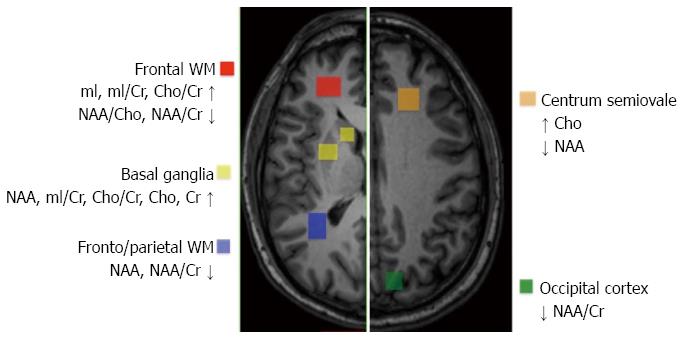
Figure 2 Quantification and localization of brain metabolite concentrations in hepatitis C virus patients exhibiting neuropsychological and neurocognitive dysfunction.
The regional distribution of proton magnetic resonance spectroscopy (1H MRS) abnormalities suggests that only cortical and subcortical telencephalic areas, but not the thalamus or posterior fossa structures, are involved in hepatitis C virus (HCV)-associated neurocognitive disorder (HCV-AND).
- Citation: Monaco S, Mariotto S, Ferrari S, Calabrese M, Zanusso G, Gajofatto A, Sansonno D, Dammacco F. Hepatitis C virus-associated neurocognitive and neuropsychiatric disorders: Advances in 2015. World J Gastroenterol 2015; 21(42): 11974-11983
- URL: https://www.wjgnet.com/1007-9327/full/v21/i42/11974.htm
- DOI: https://dx.doi.org/10.3748/wjg.v21.i42.11974


