Copyright
©The Author(s) 2015.
World J Gastroenterol. Sep 14, 2015; 21(34): 9927-9935
Published online Sep 14, 2015. doi: 10.3748/wjg.v21.i34.9927
Published online Sep 14, 2015. doi: 10.3748/wjg.v21.i34.9927
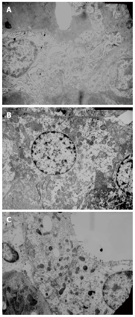
Figure 3 Histopathology of the kidney.
A: Glomeruli: Glomerular basement membrane was intact, and the pedicles of podocytes and fenestrations on endothelial cells could be clearly seen; B: Proximal tubules: The base of cuboidal epithelial cells of proximal tubules showed many plasma membrane infolds, in which many mitochondria with intact cristae were visible, and the microvilli on the luminal surface of proximal tubules were long and thick; C: Distal tubules: The base of cuboidal epithelial cells of distal tubules also contained plasma membrane infolds, in which many mitochondria were visible. The microvilli on the luminal surface of distal tubules were short and sparse.
- Citation: Wang JB, Wang HT, Li LP, Yan YC, Wang W, Liu JY, Zhao YT, Gao WS, Zhang MX. Development of a rat model of D-galactosamine/lipopolysaccharide induced hepatorenal syndrome. World J Gastroenterol 2015; 21(34): 9927-9935
- URL: https://www.wjgnet.com/1007-9327/full/v21/i34/9927.htm
- DOI: https://dx.doi.org/10.3748/wjg.v21.i34.9927


