Copyright
©The Author(s) 2015.
World J Gastroenterol. Sep 7, 2015; 21(33): 9793-9802
Published online Sep 7, 2015. doi: 10.3748/wjg.v21.i33.9793
Published online Sep 7, 2015. doi: 10.3748/wjg.v21.i33.9793
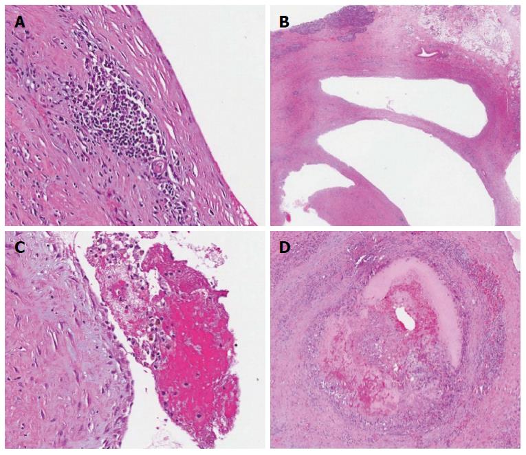
Figure 7 Histopathology.
A: The hemangioma displaced the exocrine pancreas; B: It had blood filled ectatic vessels with thin endothelium lining large cavernous spaces and aggregates of chronic inflammatory cells in the surrounding fibrous tissue; C: The lumina of the vessels contained macrophages, some containing hemosiderin; D: One vessel with organizing thrombus containing fibrin and foamy macrophages.
- Citation: Mondal U, Henkes N, Henkes D, Rosenkranz L. Cavernous hemangioma of adult pancreas: A case report and literature review. World J Gastroenterol 2015; 21(33): 9793-9802
- URL: https://www.wjgnet.com/1007-9327/full/v21/i33/9793.htm
- DOI: https://dx.doi.org/10.3748/wjg.v21.i33.9793


