Copyright
©The Author(s) 2015.
World J Gastroenterol. Sep 7, 2015; 21(33): 9774-9784
Published online Sep 7, 2015. doi: 10.3748/wjg.v21.i33.9774
Published online Sep 7, 2015. doi: 10.3748/wjg.v21.i33.9774
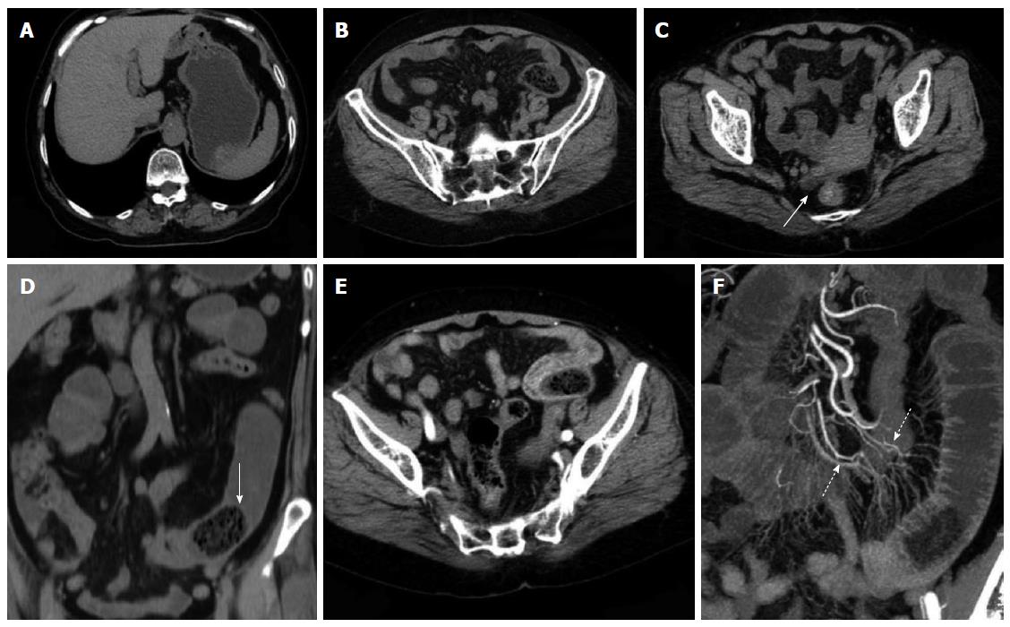
Figure 5 Representative case of a 71-year-old female with diospyrobezoar-induced small bowel obstruction.
A and B: Computed tomography (CT) planar scan image revealing oval bezoars inside the gastrointestinal anastomotic stoma and the distal end of the jejunal cavity; adjacent mesentery was slightly blurred; C: The right rear end of the uterine bottom with a small amount of seroperitoneum (white arrow); D: MPR image showing the shape of a bezoar and a linear envelope (solid arrow); E: Artery phase image displaying no thickening of the distal intestinal wall at the obstruction site but with significant enhancement; F: CTA image showing different mesenteric arteries (dotted arrow) that provide blood supply for the transitional zone and the distal end of the obstruction site.
- Citation: Wang PY, Wang X, Zhang L, Li HF, Chen L, Wang X, Wang B. Bezoar-induced small bowel obstruction: Clinical characteristics and diagnostic value of multi-slice spiral computed tomography. World J Gastroenterol 2015; 21(33): 9774-9784
- URL: https://www.wjgnet.com/1007-9327/full/v21/i33/9774.htm
- DOI: https://dx.doi.org/10.3748/wjg.v21.i33.9774


