Copyright
©The Author(s) 2015.
World J Gastroenterol. Jan 21, 2015; 21(3): 988-996
Published online Jan 21, 2015. doi: 10.3748/wjg.v21.i3.988
Published online Jan 21, 2015. doi: 10.3748/wjg.v21.i3.988
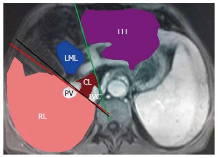
Figure 1 Outline of each liver lobe.
Right liver lobe is differentiated from the left liver lobe by the middle liver fissure (black line), and the discrimination between the left lateral liver lobe and the left medial liver lobe is provided by the liver interlobar fissure (green line) on the level of the fossa for gallbladder. The connecting line (red) between the inferior vena cava (IVC) and the right branch of the portal vein (PV) is used to distinguish the right lobe (RL) from the caudate lobe (CL). The profiles of the RL, left lateral liver lobe (LLL), left medial liver lobe (LML) and CL are drawn on the axial portal venous phase enhanced magnetic resonance imaging, and marked in pink, blue, purple and red, respectively.
- Citation: Li H, Chen TW, Li ZL, Zhang XM, Li CJ, Chen XL, Chen GW, Hu JN, Ye YQ. Albumin and magnetic resonance imaging-liver volume to identify hepatitis B-related cirrhosis and esophageal varices. World J Gastroenterol 2015; 21(3): 988-996
- URL: https://www.wjgnet.com/1007-9327/full/v21/i3/988.htm
- DOI: https://dx.doi.org/10.3748/wjg.v21.i3.988


