Copyright
©The Author(s) 2015.
World J Gastroenterol. Aug 7, 2015; 21(29): 8817-8825
Published online Aug 7, 2015. doi: 10.3748/wjg.v21.i29.8817
Published online Aug 7, 2015. doi: 10.3748/wjg.v21.i29.8817
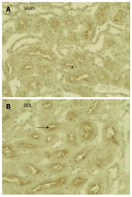
Figure 2 Immunohistochemistry for Oat5 in kidneys from Sham (A) and BDL (B) rats.
Serial sections from each rat kidney were stained using a non-commercial anti-Oat5 antibody. Oat5 labeling was associated with the apical plasma membrane domains in proximal tubule cells (arrow heads). In kidneys from BDL group there was a marked increase in Oat5 staining (arrows). These figures are representative of typical samples from four rats for each experimental group. Magnification × 200.
- Citation: Brandoni A, Torres AM. Expression of renal Oat5 and NaDC1 transporters in rats with acute biliary obstruction. World J Gastroenterol 2015; 21(29): 8817-8825
- URL: https://www.wjgnet.com/1007-9327/full/v21/i29/8817.htm
- DOI: https://dx.doi.org/10.3748/wjg.v21.i29.8817


