Copyright
©The Author(s) 2015.
World J Gastroenterol. Jul 21, 2015; 21(27): 8326-8339
Published online Jul 21, 2015. doi: 10.3748/wjg.v21.i27.8326
Published online Jul 21, 2015. doi: 10.3748/wjg.v21.i27.8326
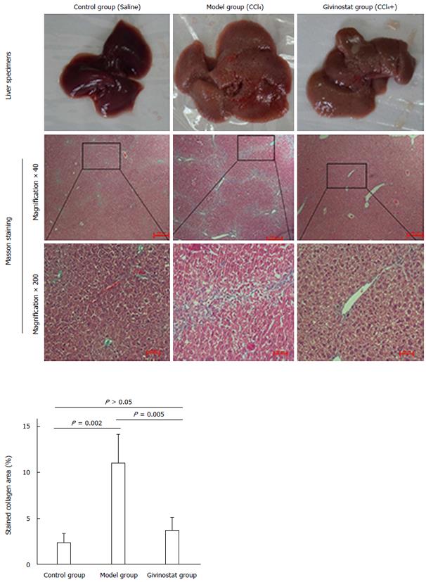
Figure 5 Pathologic changes of liver tissue in mouse models of liver fibrosis induced by CCl4 treatment.
Control group: The hepatic lobules had integrated structures, with only a small number of stained collagen fibers in the portal area. There was no obvious collagen deposition, and no degeneration, necrosis, or inflammatory cell infiltration. Model group: The normal hepatic lobules were damaged, along with the markedly increased collagen in liver tissue. The collagen fibers extended along the portal area or outwards to the inflammatory necrosis area, forming fiber spacing with varied thickness. Pseudolobules were visible. Givinostat group: compared with the model group, the structure of liver tissue was markedly improved, along with the obviously decreased collagen deposition. Collagen area percent was significantly reduced in givinostat group compared to the model group (P = 0.005).
- Citation: Wang YG, Xu L, Wang T, Wei J, Meng WY, Wang N, Shi M. Givinostat inhibition of hepatic stellate cell proliferation and protein acetylation. World J Gastroenterol 2015; 21(27): 8326-8339
- URL: https://www.wjgnet.com/1007-9327/full/v21/i27/8326.htm
- DOI: https://dx.doi.org/10.3748/wjg.v21.i27.8326


