Copyright
©The Author(s) 2015.
World J Gastroenterol. Jul 14, 2015; 21(26): 8148-8155
Published online Jul 14, 2015. doi: 10.3748/wjg.v21.i26.8148
Published online Jul 14, 2015. doi: 10.3748/wjg.v21.i26.8148
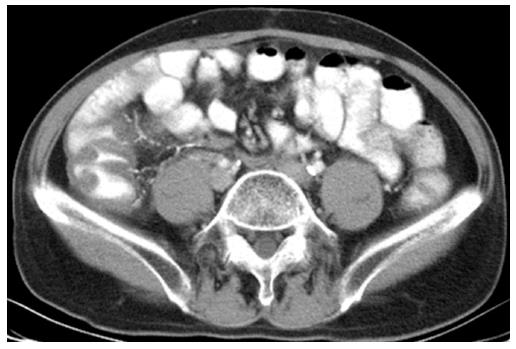
Figure 3 Intravenous contrast enhanced computed tomography with oral contrast ingestion.
A 59-year-old male came to emergency room with fever and tenderness at right lower abdomen. The contrast enhanced computed tomography shows wall thickening at the ascending colon and calcifications at straight veins, marginal veins and main branch of mesenteric vein, suggestive of phlebosclerotic colitis.
- Citation: Yen TS, Liu CA, Chiu NC, Chiou YY, Chou YH, Chang CY. Relationship between severity of venous calcifications and symptoms of phlebosclerotic colitis. World J Gastroenterol 2015; 21(26): 8148-8155
- URL: https://www.wjgnet.com/1007-9327/full/v21/i26/8148.htm
- DOI: https://dx.doi.org/10.3748/wjg.v21.i26.8148


