Copyright
©The Author(s) 2015.
World J Gastroenterol. Jul 14, 2015; 21(26): 8148-8155
Published online Jul 14, 2015. doi: 10.3748/wjg.v21.i26.8148
Published online Jul 14, 2015. doi: 10.3748/wjg.v21.i26.8148
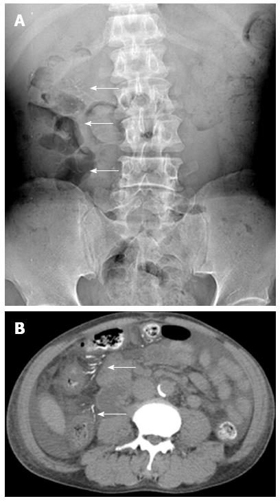
Figure 2 Kidney-ureter-bladder radiography and non-contrast enhanced computed tomography.
A 56-year-old male suffered from right upper quadrant abdominal pain and fever. A: The kidney-ureter-bladder radiography showed threadlike calcifications (arrows) at right abdomen; B: The non-contrast enhanced computed tomography study revealed calcifications (arrows) at tributaries of mesenteric vein and wall thickening of the ascending colon. The diagnosis is phlebosclerotic colitis with active episode.
- Citation: Yen TS, Liu CA, Chiu NC, Chiou YY, Chou YH, Chang CY. Relationship between severity of venous calcifications and symptoms of phlebosclerotic colitis. World J Gastroenterol 2015; 21(26): 8148-8155
- URL: https://www.wjgnet.com/1007-9327/full/v21/i26/8148.htm
- DOI: https://dx.doi.org/10.3748/wjg.v21.i26.8148


