Copyright
©The Author(s) 2015.
World J Gastroenterol. Jul 7, 2015; 21(25): 7916-7920
Published online Jul 7, 2015. doi: 10.3748/wjg.v21.i25.7916
Published online Jul 7, 2015. doi: 10.3748/wjg.v21.i25.7916
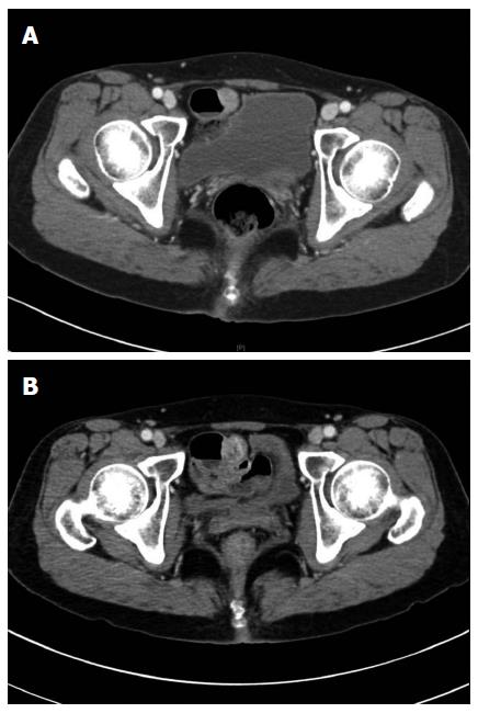
Figure 1 Abdominopelvic computed tomography in 2011 and B: 2014.
A: Contrast enhanced computed tomography (CT) scan shows 1.7 cm sized enhancing mass arising in small bowel; B: Follow up CT shows a slightly increased size to 2.2 cm and new appearance of low density areas in the mass suggests necrotic change.
- Citation: Lee SH, Kim WY, Hwang DY, Han HS. Intraductal papillary mucinous neoplasm of the ileal heterotopic pancreas in a patient with hereditary non-polyposis colorectal cancer: A case report. World J Gastroenterol 2015; 21(25): 7916-7920
- URL: https://www.wjgnet.com/1007-9327/full/v21/i25/7916.htm
- DOI: https://dx.doi.org/10.3748/wjg.v21.i25.7916


