Copyright
©The Author(s) 2015.
World J Gastroenterol. Jun 21, 2015; 21(23): 7335-7342
Published online Jun 21, 2015. doi: 10.3748/wjg.v21.i23.7335
Published online Jun 21, 2015. doi: 10.3748/wjg.v21.i23.7335
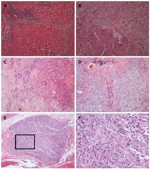
Figure 2 Microscopic appearance of the specimen (hematoxylin and eosin staining).
A: Tumor-adjacent liver parenchyma showed portal cirrhosis (magnificaton × 100); B: A transitional zone could be defined between the carcinomatous (lower left) and sarcomatous components (upper right). There were intermingled cells of the two elements (magnificaton × 100); C: Foci of cholangiocellular carcinoma with various grades of differentiation (magnificaton × 100); D: Neoplastic osteoid and chondroid formation was observed in the sarcoma element (magnificaton × 100); E: Metastasis nodule was composed of undifferentiated sarcoma cells with deeply stained nuclei (magnificaton × 40); F: Higher-magnification view of the squared part in panel E.
- Citation: Xiang S, Chen YF, Guan Y, Chen XP. Primary combined hepatocellular-cholangiocellular sarcoma: An unusual case. World J Gastroenterol 2015; 21(23): 7335-7342
- URL: https://www.wjgnet.com/1007-9327/full/v21/i23/7335.htm
- DOI: https://dx.doi.org/10.3748/wjg.v21.i23.7335


