Copyright
©The Author(s) 2015.
World J Gastroenterol. Jun 14, 2015; 21(22): 7052-7058
Published online Jun 14, 2015. doi: 10.3748/wjg.v21.i22.7052
Published online Jun 14, 2015. doi: 10.3748/wjg.v21.i22.7052
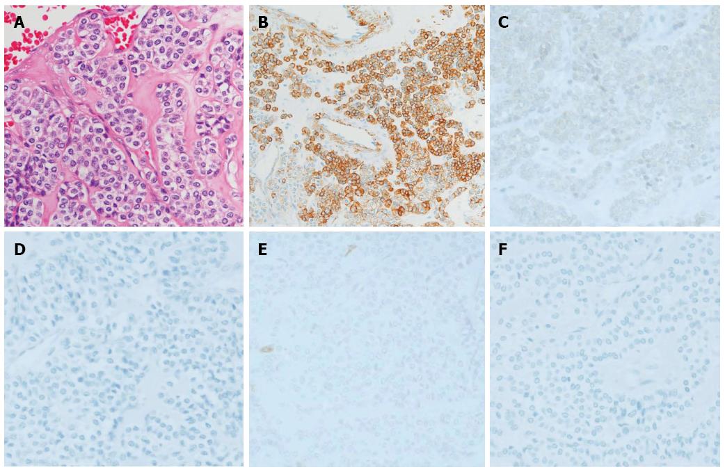
Figure 4 Histology and Immunohistochemical analysis of resected specimen was consistent with the endoscopic ultrasound-guided fine-needle aspiration biopsy pathology results.
A: Hematoxylin and eosin stain (× 400); B: Muscle actin; C: Synaptophysin; D: Chromogranin; E: c-kit; F: Desmin (× 400).
- Citation: Kato S, Kikuchi K, Chinen K, Murakami T, Kunishima F. Diagnostic utility of endoscopic ultrasound-guided fine-needle aspiration biopsy for glomus tumor of the stomach. World J Gastroenterol 2015; 21(22): 7052-7058
- URL: https://www.wjgnet.com/1007-9327/full/v21/i22/7052.htm
- DOI: https://dx.doi.org/10.3748/wjg.v21.i22.7052


