Copyright
©The Author(s) 2015.
World J Gastroenterol. Jun 14, 2015; 21(22): 6850-6860
Published online Jun 14, 2015. doi: 10.3748/wjg.v21.i22.6850
Published online Jun 14, 2015. doi: 10.3748/wjg.v21.i22.6850
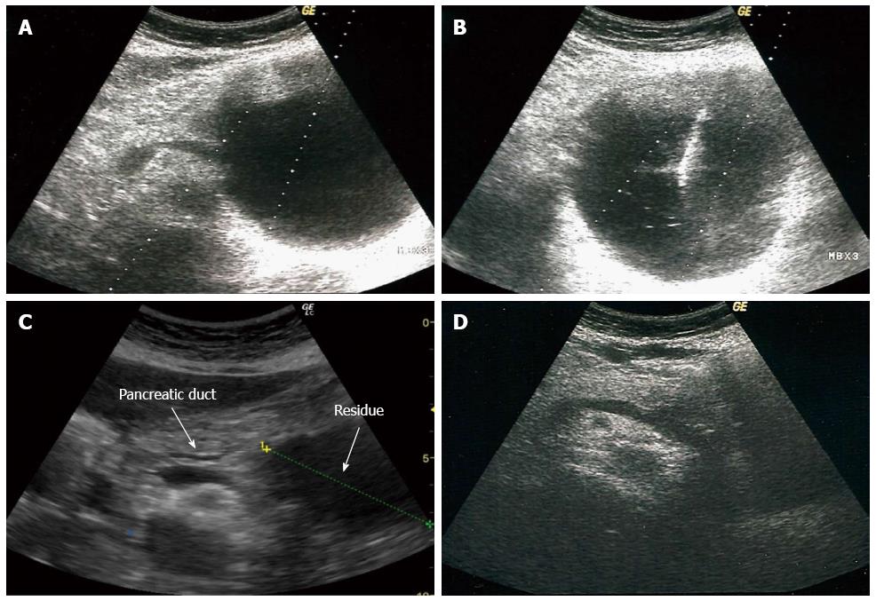
Figure 1 Appearance on ultrasound of a pancreatic pseudocyst before, during and after treatment.
A: Appearance on ultrasound of a PPC in the tail of pancreas before treatment; B: Insertion of a catheter into the PPC; C: Residue of PPC with suspected PPC-PD communication (marked by arrows) immediately after the procedure; D: The appearance of the pancreas several months after the procedure. PPC: Pancreatic pseudocyst; PD: Pancreatic duct.
- Citation: Zerem E, Hauser G, Loga-Zec S, Kunosić S, Jovanović P, Crnkić D. Minimally invasive treatment of pancreatic pseudocysts. World J Gastroenterol 2015; 21(22): 6850-6860
- URL: https://www.wjgnet.com/1007-9327/full/v21/i22/6850.htm
- DOI: https://dx.doi.org/10.3748/wjg.v21.i22.6850


