Copyright
©The Author(s) 2015.
World J Gastroenterol. Jun 7, 2015; 21(21): 6591-6603
Published online Jun 7, 2015. doi: 10.3748/wjg.v21.i21.6591
Published online Jun 7, 2015. doi: 10.3748/wjg.v21.i21.6591
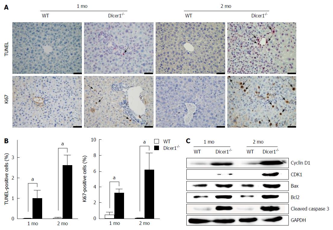
Figure 4 Dicer1-/- liver displays increased hepatocyte death and compensatory proliferation.
A: Significant hepatocyte apoptosis and proliferation in the Dicer1-deficient liver (Scale bar: 100 μm); B: Quantification of hepatocyte apoptosis (TUNEL staining) and proliferation (Ki67 immunohistochemistry) in Dicer1-deficient liver (mean ± SD; n = 4-5), aP < 0.05 vs wild-type mice; C: Western blot analysis of the liver tissue lysates with indicated antibodies. GAPDH was used as an internal control.
- Citation: Lu XF, Zhou YJ, Zhang L, Ji HJ, Li L, Shi YJ, Bu H. Loss of Dicer1 impairs hepatocyte survival and leads to chronic inflammation and progenitor cell activation. World J Gastroenterol 2015; 21(21): 6591-6603
- URL: https://www.wjgnet.com/1007-9327/full/v21/i21/6591.htm
- DOI: https://dx.doi.org/10.3748/wjg.v21.i21.6591


