Copyright
©The Author(s) 2015.
World J Gastroenterol. Jan 14, 2015; 21(2): 675-687
Published online Jan 14, 2015. doi: 10.3748/wjg.v21.i2.675
Published online Jan 14, 2015. doi: 10.3748/wjg.v21.i2.675
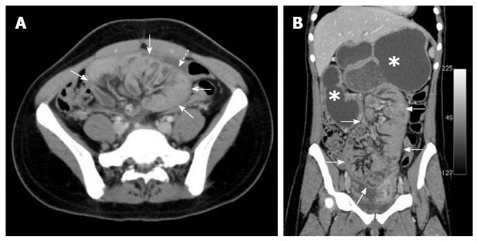
Figure 4 Contrast-enhanced abdominal computed tomography[24].
Small intestinal loops are encased in a sac of thick peritoneal membrane (continuous arrows) with a small volume of peritoneal liquid effusion (discontinuous arrow). Gastroduodenal distension is also present (asterisks). A: Axial slice; B: Multiplanar coronal reconstruction.
- Citation: Akbulut S. Accurate definition and management of idiopathic sclerosing encapsulating peritonitis. World J Gastroenterol 2015; 21(2): 675-687
- URL: https://www.wjgnet.com/1007-9327/full/v21/i2/675.htm
- DOI: https://dx.doi.org/10.3748/wjg.v21.i2.675


