Copyright
©The Author(s) 2015.
World J Gastroenterol. Jan 14, 2015; 21(2): 517-524
Published online Jan 14, 2015. doi: 10.3748/wjg.v21.i2.517
Published online Jan 14, 2015. doi: 10.3748/wjg.v21.i2.517
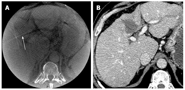
Figure 1 Illustration of a poor quality cone beam computed tomography image obtained in a 65-year-old man with hepatocellular carcinoma on hepatitis C virus cirrhosis background.
A: Visibility of the ablation zone is poor (arrow); B: Post-ablation multi-detector computed tomography performed at 6 wk showed complete ablation.
- Citation: Abdel-Rehim M, Ronot M, Sibert A, Vilgrain V. Assessment of liver ablation using cone beam computed tomography. World J Gastroenterol 2015; 21(2): 517-524
- URL: https://www.wjgnet.com/1007-9327/full/v21/i2/517.htm
- DOI: https://dx.doi.org/10.3748/wjg.v21.i2.517


