Copyright
©The Author(s) 2015.
World J Gastroenterol. May 21, 2015; 21(19): 6088-6096
Published online May 21, 2015. doi: 10.3748/wjg.v21.i19.6088
Published online May 21, 2015. doi: 10.3748/wjg.v21.i19.6088
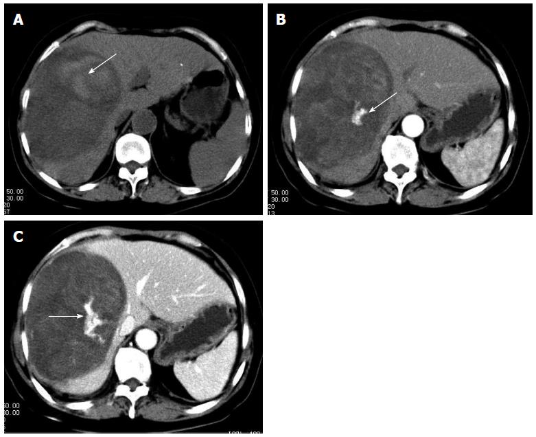
Figure 5 Computed tomography scan.
A: Plain computed tomography scan reveals an oval shaped heterogeneous hypoattenuating mass with hyperattenuating foci (arrow) which suggests hemorrhage; B: Dominant mass lesion shows heterogenous enhancement with hyperattenuating enhanced foci (arrow), which indicate vasculature, in arterial phase; C: Dominant mass shows progressive enhancement and hyperattenuating enhanced foci enlarged in portal phase (arrows).
- Citation: Zhu YP, Chen YM, Matro E, Chen RB, Jiang ZN, Mou YP, Hu HJ, Huang CJ, Wang GY. Primary hepatic angiosarcoma: A report of two cases and literature review. World J Gastroenterol 2015; 21(19): 6088-6096
- URL: https://www.wjgnet.com/1007-9327/full/v21/i19/6088.htm
- DOI: https://dx.doi.org/10.3748/wjg.v21.i19.6088


