Copyright
©The Author(s) 2015.
World J Gastroenterol. May 14, 2015; 21(18): 5635-5640
Published online May 14, 2015. doi: 10.3748/wjg.v21.i18.5635
Published online May 14, 2015. doi: 10.3748/wjg.v21.i18.5635
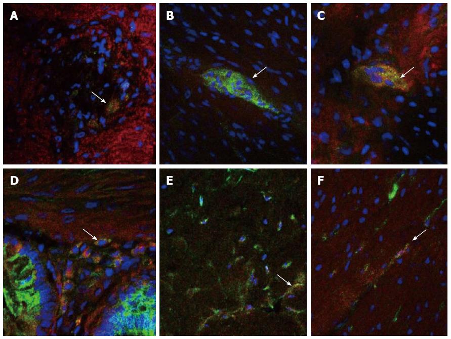
Figure 1 Immunofluorescent staining in normal control tissue of HCN2 channels (green), co-localised (arrows) with neuronal marker HuC/D (red) (A) and c-kit (red) (D), HCN3 channels (red) co-localised with PGP9.
5 (green) (B), and TMEM (green) (E), and HCN4 channels (green) co-localised with HuC/D (red) (C) and c-kit (red) (F). Nuclei were stained with DAPI (blue).
- Citation: O’Donnell AM, Coyle D, Puri P. Decreased expression of hyperpolarisation-activated cyclic nucleotide-gated channel 3 in Hirschsprung’s disease. World J Gastroenterol 2015; 21(18): 5635-5640
- URL: https://www.wjgnet.com/1007-9327/full/v21/i18/5635.htm
- DOI: https://dx.doi.org/10.3748/wjg.v21.i18.5635


