Copyright
©The Author(s) 2015.
World J Gastroenterol. Apr 28, 2015; 21(16): 4883-4893
Published online Apr 28, 2015. doi: 10.3748/wjg.v21.i16.4883
Published online Apr 28, 2015. doi: 10.3748/wjg.v21.i16.4883
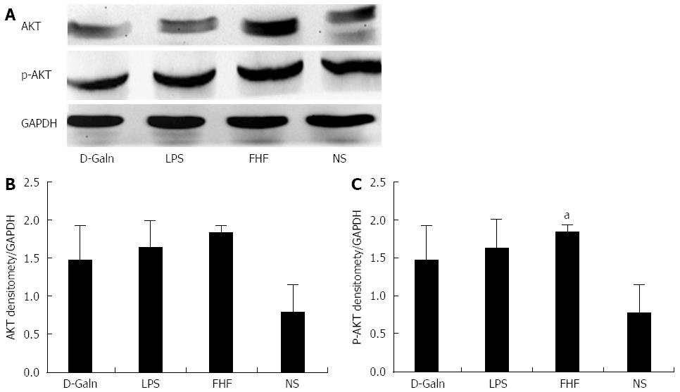
Figure 8 Intestinal AKT and phosphorylated-AKT protein expression.
A: AKT and p-AKT protein expression was detected by Western blot; B and C: Densitometric analysis using the Image-Pro software. The ratio of the absorbance of AKT/absorbance of GAPDH in the fulminant hepatic failure (FHF) group was not significantly different compared with those of normal saline (NS), lipopolysaccharide (LPS) and D-galactosamine (D-Galn) groups. The ratio of the absorbance of phosphorylated-AKT (p-AKT)/absorbance of GAPDH in the FHF group was notably increased compared with that of NS group, but was not significantly different compared with those of the LPS and D-Galn groups (aP < 0.05 vs the NS group, one-way ANOVA with Dunnett test; n≥ 15).
- Citation: Cao X, Liu M, Wang P, Liu DY. Intestinal dendritic cells change in number in fulminant hepatic failure. World J Gastroenterol 2015; 21(16): 4883-4893
- URL: https://www.wjgnet.com/1007-9327/full/v21/i16/4883.htm
- DOI: https://dx.doi.org/10.3748/wjg.v21.i16.4883


