Copyright
©The Author(s) 2015.
World J Gastroenterol. Apr 7, 2015; 21(13): 4069-4077
Published online Apr 7, 2015. doi: 10.3748/wjg.v21.i13.4069
Published online Apr 7, 2015. doi: 10.3748/wjg.v21.i13.4069
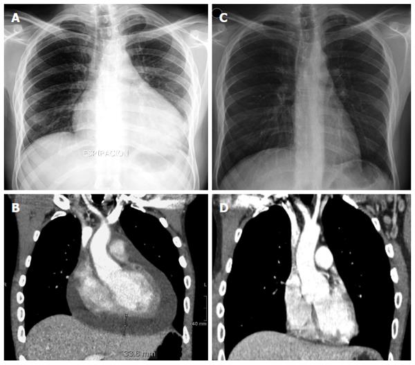
Figure 1 Radiograph of the chest and computed tomography scan before and after mesalazine suspension.
A: Chest radiograph showing an enlarged cardiac silhouette due to a cardiac effusion during mesalazine treatment; B: Computed tomography (CT) scan of the chest revealing cardiomegaly due to a large pericardial effusion (maximum width of 33.6 mm) during mesalazine therapy; C: Normal chest radiograph after mesalazine withdrawal; D: CT scan of the chest showing a complete resolution of the pericardial effusion after suspension of mesalazine.
- Citation: Ferrusquía J, Pérez-Martínez I, Gómez de la Torre R, Fernández-Almira ML, de Francisco R, Rodrigo L, Riestra S. Gastroenterology case report of mesalazine-induced cardiopulmonary hypersensitivity. World J Gastroenterol 2015; 21(13): 4069-4077
- URL: https://www.wjgnet.com/1007-9327/full/v21/i13/4069.htm
- DOI: https://dx.doi.org/10.3748/wjg.v21.i13.4069


