Copyright
©The Author(s) 2015.
World J Gastroenterol. Mar 28, 2015; 21(12): 3499-3508
Published online Mar 28, 2015. doi: 10.3748/wjg.v21.i12.3499
Published online Mar 28, 2015. doi: 10.3748/wjg.v21.i12.3499
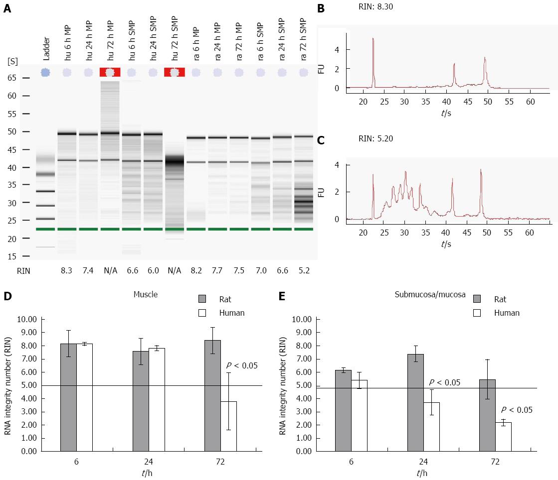
Figure 1 Time course of degradation effect on muscle and submucosal/mucosal human and rat tissue.
Human and rat gut samples were stored in MEM-Hepes on ice and separated in smooth muscle and submucosal/mucosal layer (muscle, mucosa) at different points in time. RNA from the tissues was isolated and degradation (RIN) of the different samples was examined. A: The figure shows a typical gel for human and rodent samples with different integrities. First the ladder, followed by 3 human muscle samples (hu MP), another 3 samples from human mucous layer (hu SMP) all from different points in time. The last 6 samples were comparable samples from rat (ra); B and C: Typical electrophoretic RNA measurements obtained from an Agilent 2100 bioanalyzer displaying different RIN values for intact (8 and 3 respectively; B) and partly degraded RNA (5 and 2 respectively; C); RIN values from both rat and human tissues from different compartment and time points are depicted in (D) and (E). Data are presented as the mean ± SE. n = 10.
- Citation: Heumüller-Klug S, Sticht C, Kaiser K, Wink E, Hagl C, Wessel L, Schäfer KH. Degradation of intestinal mRNA: A matter of treatment. World J Gastroenterol 2015; 21(12): 3499-3508
- URL: https://www.wjgnet.com/1007-9327/full/v21/i12/3499.htm
- DOI: https://dx.doi.org/10.3748/wjg.v21.i12.3499


