Copyright
©The Author(s) 2015.
World J Gastroenterol. Mar 21, 2015; 21(11): 3429-3434
Published online Mar 21, 2015. doi: 10.3748/wjg.v21.i11.3429
Published online Mar 21, 2015. doi: 10.3748/wjg.v21.i11.3429
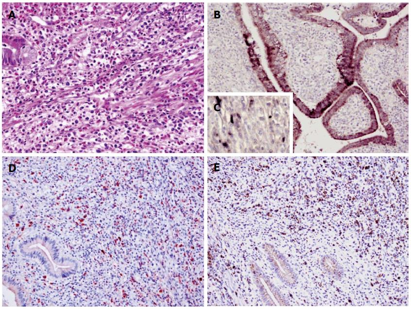
Figure 4 Histological findings of the resected gallbladder specimen.
HE staining shows abundant infiltration of lymphoplasmacytes and eosinophils (A). Immunostaining shows Helicobacter pylori in the epithelium (B) or mesenchymal cells (C), IgG4-positive (D) or IgG-positive plasma cells (E) in the resected gallbladder sections of the patient. Original magnification × 400 (A and C); × 200 (B, D and E).
-
Citation: Li M, Zhou Q, Yang K, Brigstock DR, Zhang L, Xiu M, Sun L, Gao RP. Rare case of
Helicobacter pylori -positive multiorgan IgG4-related disease and gastric cancer. World J Gastroenterol 2015; 21(11): 3429-3434 - URL: https://www.wjgnet.com/1007-9327/full/v21/i11/3429.htm
- DOI: https://dx.doi.org/10.3748/wjg.v21.i11.3429


