Copyright
©The Author(s) 2015.
World J Gastroenterol. Mar 21, 2015; 21(11): 3380-3387
Published online Mar 21, 2015. doi: 10.3748/wjg.v21.i11.3380
Published online Mar 21, 2015. doi: 10.3748/wjg.v21.i11.3380
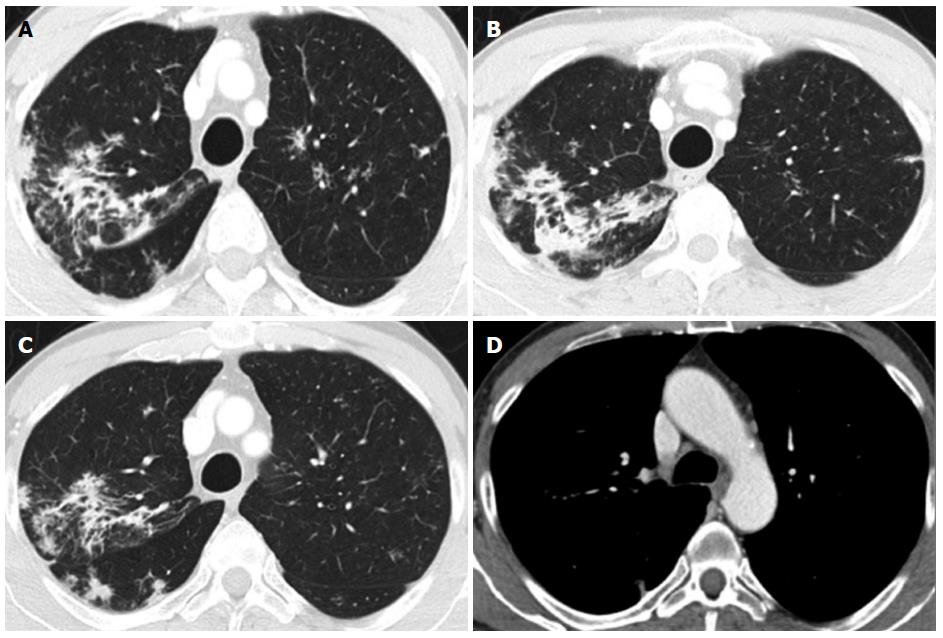
Figure 2 Chest computed tomography performed after colonoscopy for whole-body staging.
A and B: Asymmetric discrete airspace consolidation with air bronchograms in the right upper lobe; C: Numerous micronodular opacities are present in both upper pulmonary lobes (also seen in panel A) with distribution in the peribronchovascular interstitium. These findings showed no significant change after four weeks of antibiotic therapy; D: No pathologic hilar or mediastinal lymph node enlargement was observed.
- Citation: Erra P, Crusco S, Nugnes L, Pollio AM, Di Pilla G, Biondi G, Vigliardi G. Colonic sarcoidosis: Unusual onset of a systemic disease. World J Gastroenterol 2015; 21(11): 3380-3387
- URL: https://www.wjgnet.com/1007-9327/full/v21/i11/3380.htm
- DOI: https://dx.doi.org/10.3748/wjg.v21.i11.3380


