Copyright
©The Author(s) 2015.
World J Gastroenterol. Mar 14, 2015; 21(10): 2988-2996
Published online Mar 14, 2015. doi: 10.3748/wjg.v21.i10.2988
Published online Mar 14, 2015. doi: 10.3748/wjg.v21.i10.2988
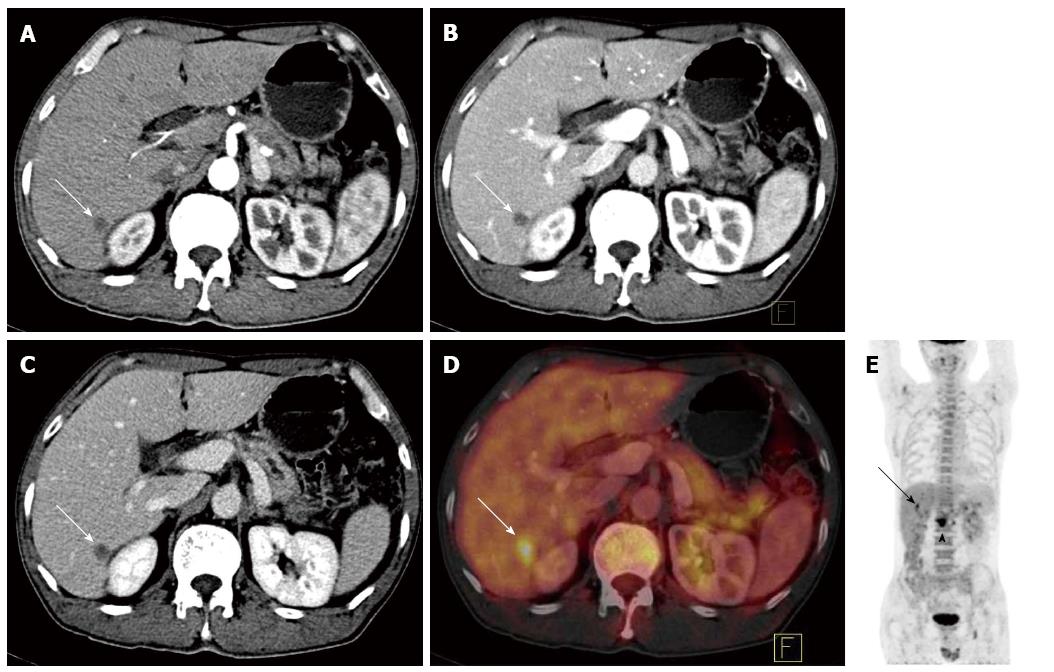
Figure 3 A 49-year-old pancreatic cancer patient.
A-C: CECT images at the arterial phase, pancreatic parenchymal phase, and delayed phase. A tiny, round, low density, 9 mm lesion was found at the posterior right liver lobe (arrow). This lesion was diagnosed as a hepatic cyst by CECT imaging because no enhancement was found in the lesion; D: A PET/CECT fusion image showed increased FDG uptake at the lesion with an SUVmax of 5.2. This lesion was diagnosed as liver metastasis from pancreatic cancer with PET/CECT fusion images, which was confirmed by biopsy; E: PET image for the entire body. A high metabolic mass was found at the pancreatic head (triangle). PET/CT: Positron emission tomography/computed tomography; CECT: Contrast-enhanced CT; FDG: Fluorodeoxyglucose.
- Citation: Zhang J, Zuo CJ, Jia NY, Wang JH, Hu SP, Yu ZF, Zheng Y, Zhang AY, Feng XY. Cross-modality PET/CT and contrast-enhanced CT imaging for pancreatic cancer. World J Gastroenterol 2015; 21(10): 2988-2996
- URL: https://www.wjgnet.com/1007-9327/full/v21/i10/2988.htm
- DOI: https://dx.doi.org/10.3748/wjg.v21.i10.2988


