Copyright
©The Author(s) 2015.
World J Gastroenterol. Jan 7, 2015; 21(1): 60-83
Published online Jan 7, 2015. doi: 10.3748/wjg.v21.i1.60
Published online Jan 7, 2015. doi: 10.3748/wjg.v21.i1.60
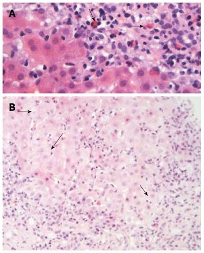
Figure 4 Liver histology in autoimmune hepatitis (continued).
A: In some cases of autoimmune hepatitis, “numerous” eosinophils or eosinophils as a minor component of the inflammatory infiltrate can be seen (arrows; hematoxylin and eosin staining). This is an interesting and useful observation, since this finding is evaluated falsely out of the histopathological context of autoimmune hepatitis, and therefore, could make the differential diagnosis from drug-induced hepatitis more problematic; B: Interface hepatitis with hydropic change of involved hepatocytes (arrows; hematoxylin and eosin staining). Note the presence of adjacent parenchymal collapse, alternatively multiacinar necrosis, which has been considered as one of the most distinguishing morphological features of autoimmune hepatitis since its presence in the appropriate clinicopathological context is supportive of the diagnosis.
- Citation: Gatselis NK, Zachou K, Koukoulis GK, Dalekos GN. Autoimmune hepatitis, one disease with many faces: Etiopathogenetic, clinico-laboratory and histological characteristics. World J Gastroenterol 2015; 21(1): 60-83
- URL: https://www.wjgnet.com/1007-9327/full/v21/i1/60.htm
- DOI: https://dx.doi.org/10.3748/wjg.v21.i1.60


