Copyright
©2014 Baishideng Publishing Group Co.
World J Gastroenterol. Mar 7, 2014; 20(9): 2420-2425
Published online Mar 7, 2014. doi: 10.3748/wjg.v20.i9.2420
Published online Mar 7, 2014. doi: 10.3748/wjg.v20.i9.2420
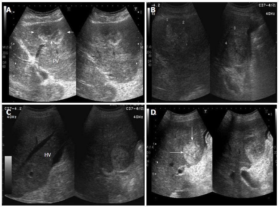
Figure 1 Abdominal ultrasound showing multiple hyperechoic or mixed echoic tumors in the bilateral lobes of liver (arrows).
Minimal ascites was also detected. A: One mixed echoic tumor (arrows) at segment 3, left portal vein (PV); B: One mixed echoic tumor (marked) at segment 5; C: One hyperechoic tumor at segment 5, right hepatic vein (HV), minimal ascites also detected; D: One hyperechoic tumor (arrows) at segment 5 near the main portal trunk.
- Citation: Yu CY, Chang LC, Chen LW, Lee TS, Chien RN, Hsieh MF, Chiang KC. Peliosis hepatis complicated by portal hypertension following renal transplantation. World J Gastroenterol 2014; 20(9): 2420-2425
- URL: https://www.wjgnet.com/1007-9327/full/v20/i9/2420.htm
- DOI: https://dx.doi.org/10.3748/wjg.v20.i9.2420


