Copyright
©2014 Baishideng Publishing Group Co.
World J Gastroenterol. Mar 7, 2014; 20(9): 2403-2411
Published online Mar 7, 2014. doi: 10.3748/wjg.v20.i9.2403
Published online Mar 7, 2014. doi: 10.3748/wjg.v20.i9.2403
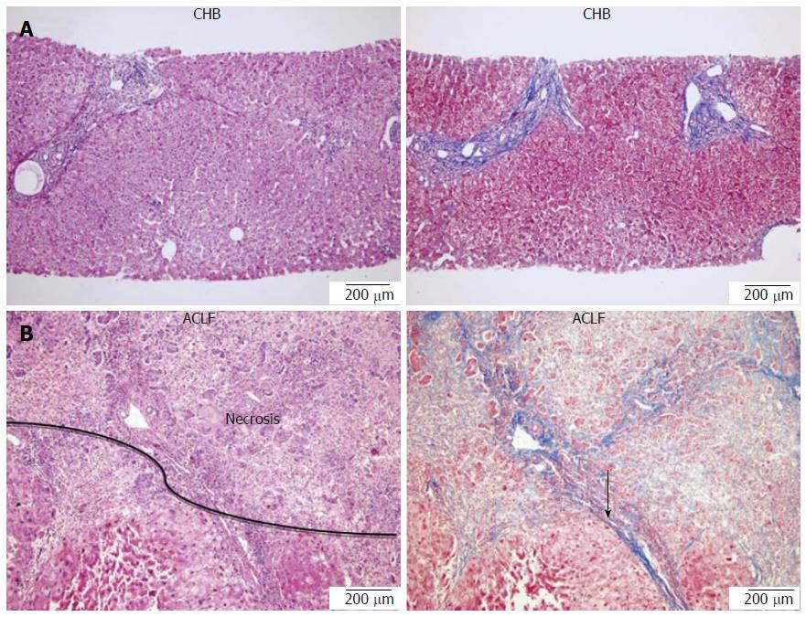
Figure 1 Histology of tissue sample for chronic hepatitis B and and acute-on-chronic liver failure in representative patients.
A: Chronic hepatitis B (CHB, mild): Mild enlarged portal tract is infiltrated by lymphocytes without interface hepatitis. Spot necrosis is in the lobule (left, HE, × 100). Portal tract shows mild fibrosis (right, Masson trichrome); B: Acute-on-chronic liver failure (ACLF): Massive necrosis of parenchyma with cirrhotic nodule remaining (left, HE, × 100). Cirrhotic nodule is surrounded by fibrous tissue (arrow) (right, Masson trichrome).
- Citation: Zheng SJ, Liu S, Liu M, McCrae MA, Li JF, Han YP, Xu CH, Ren F, Chen Y, Duan ZP. Prognostic value of M30/M65 for outcome of hepatitis B virus-related acute-on-chronic liver failure. World J Gastroenterol 2014; 20(9): 2403-2411
- URL: https://www.wjgnet.com/1007-9327/full/v20/i9/2403.htm
- DOI: https://dx.doi.org/10.3748/wjg.v20.i9.2403


