Copyright
©2014 Baishideng Publishing Group Co.
World J Gastroenterol. Feb 14, 2014; 20(6): 1630-1634
Published online Feb 14, 2014. doi: 10.3748/wjg.v20.i6.1630
Published online Feb 14, 2014. doi: 10.3748/wjg.v20.i6.1630
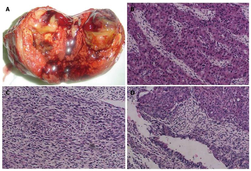
Figure 4 Pathologic findings of the primary hepatic carcinosarcoma (hematoxylin and eosin, original magnification × 200).
A: A section of the gross specimen showing a well-demarcated, peripheral nodular solid mass with a central area of necrosis and hemorrhage; B: Light microscopy showing moderate hepatocellular carcinoma; C: Stromal sarcoma components are comprised of spindle cells; D: Atypical spindle and epithelial cells are seen, which are consistent with hepatic carcinosarcoma.
- Citation: Liu LP, Yu XL, Liang P, Dong BW. Characterization of primary hepatic carcinosarcoma by contrast-enhanced ultrasonography: A case report. World J Gastroenterol 2014; 20(6): 1630-1634
- URL: https://www.wjgnet.com/1007-9327/full/v20/i6/1630.htm
- DOI: https://dx.doi.org/10.3748/wjg.v20.i6.1630


