Copyright
©2014 Baishideng Publishing Group Co.
World J Gastroenterol. Feb 14, 2014; 20(6): 1630-1634
Published online Feb 14, 2014. doi: 10.3748/wjg.v20.i6.1630
Published online Feb 14, 2014. doi: 10.3748/wjg.v20.i6.1630
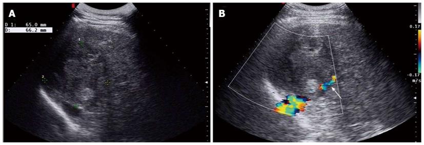
Figure 1 Conventional ultrasonography of a primary hepatic carcinosarcoma.
A: Gray-scale ultrasonography showed an ovoid heterogeneous echogenic mass in the right liver; B: Color doppler imaging showed the right hepatic vein compressed by the mass (arrow).
- Citation: Liu LP, Yu XL, Liang P, Dong BW. Characterization of primary hepatic carcinosarcoma by contrast-enhanced ultrasonography: A case report. World J Gastroenterol 2014; 20(6): 1630-1634
- URL: https://www.wjgnet.com/1007-9327/full/v20/i6/1630.htm
- DOI: https://dx.doi.org/10.3748/wjg.v20.i6.1630


