Copyright
©2014 Baishideng Publishing Group Co.
World J Gastroenterol. Feb 14, 2014; 20(6): 1493-1502
Published online Feb 14, 2014. doi: 10.3748/wjg.v20.i6.1493
Published online Feb 14, 2014. doi: 10.3748/wjg.v20.i6.1493
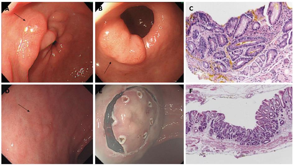
Figure 2 Metachronous gastric cancer that developed after 6 years of Helicobacter pylori eradication.
A 61 year-old South Korean man visited because of epigastric discomfort in March 2007. A: Initial endoscopic finding. Several raised erosions with central ulceration (arrow) were evident. Since H. pylori infection was found by gastric biopsy, eradication was achieved using amoxicillin (1 g), clarithromycin (500 mg), and a a standard dose of proton pump inhibitor twice daily for 7 d; B: Endoscopic finding after 2 years. In June 2009, a gastric adenoma near the pylorus (arrow) was diagnosed by endoscopic biopsy. Complete endoscopic resection was performed; C: Immunohistochemical staining of the resected specimen. Ki-67 staining was positive in the adenoma (Ki-67 stain, x 400); D: Endoscopic finding after 6 years. In January 2013, a slightly depressed lesion was evident on the lesser curvature side of the mid-antrum (arrow); E: Endoscopic submucosal dissection. The lesion was resected since the endoscopic biopsy revealed an adenocarcinoma; F: Pathological finding of the resected specimen. Early gastric cancer type IIc of Lauren’s intestinal-type, moderately-differentiated, tubular adenocarcinoma was diagnosed. The tumor size was 8.0 mm x 6.0 mm x 1.0 mm, and the depth of invasion was limited to the lamina propria (pT1a). Resection margins were free from carcinoma.
-
Citation: Lee SY. Current progress toward eradicating
Helicobacter pylori in East Asian countries: Differences in the 2013 revised guidelines between China, Japan, and South Korea. World J Gastroenterol 2014; 20(6): 1493-1502 - URL: https://www.wjgnet.com/1007-9327/full/v20/i6/1493.htm
- DOI: https://dx.doi.org/10.3748/wjg.v20.i6.1493


