Copyright
©2014 Baishideng Publishing Group Co.
World J Gastroenterol. Feb 14, 2014; 20(6): 1402-1423
Published online Feb 14, 2014. doi: 10.3748/wjg.v20.i6.1402
Published online Feb 14, 2014. doi: 10.3748/wjg.v20.i6.1402
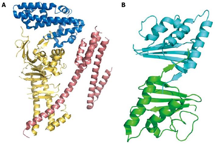
Figure 2 Cartoon model of Cag proteins.
A: The N-terminal portion of cytotoxic-associated genes, CagA, the effector protein injected into the host cell through type IV secretion system. The three domains (residues 24-221, blue; 303-644, yellow; 645-824, salmon; coordinates from PDB 4DVY) are shown in different colors; B: CagD dimer. The two monomers are linked together by a disulfide bridge between the two C-terminal β-strands. Coordinates from PDB 3CWX.
-
Citation: Zanotti G, Cendron L. Structural and functional aspects of the
Helicobacter pylori secretome. World J Gastroenterol 2014; 20(6): 1402-1423 - URL: https://www.wjgnet.com/1007-9327/full/v20/i6/1402.htm
- DOI: https://dx.doi.org/10.3748/wjg.v20.i6.1402


