Copyright
©2014 Baishideng Publishing Group Inc.
World J Gastroenterol. Dec 28, 2014; 20(48): 18495-18502
Published online Dec 28, 2014. doi: 10.3748/wjg.v20.i48.18495
Published online Dec 28, 2014. doi: 10.3748/wjg.v20.i48.18495
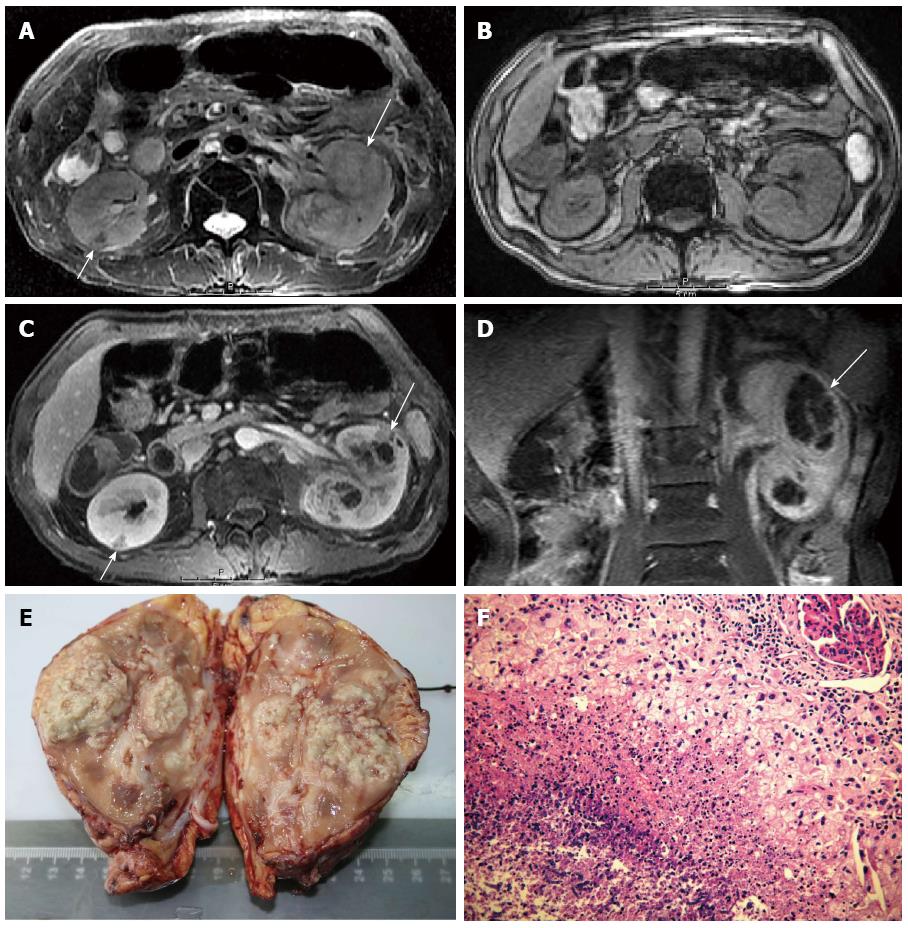
Figure 2 Renal aspergillosis in a 53-year-old allogenetic orthotropic liver transplantation recipient (Case two).
A: Fat-suppressed T2WI, which shows two large foci in the left kidney and one small focus in the right. The large foci in the left kidney present slightly high signal centers with slightly low signal walls and separations (long arrow). The wedge-shaped small focus in the right renal cortex presents low signal intensity (short arrow); B: Unenhanced out-of-phase dual-echo chemical shift T1WI; C: Contrast-enhanced T1WI on the parenchymal nephrographic phase. All foci present iso- or slightly low signal intensity and ill-defined margins on the unenhanced T1WI. On the contrast-enhanced T1WI, the large foci present enhanced irregular abscess walls and separations with unenhanced necrotic areas (long arrow); and the small focus (short arrow) presents decreased enhancement; D: Contrast-enhanced coronal T1WI, on which the large focus is typically wedge-shaped (long arrow); E: Macroscopic aspect of the left kidney with multiple foci in it; F: Pathological findings under light microscope (HE, × 40), which shows amounts of fungal hyphae and spores in the necrosis. RAsp: Renal aspergillosis; T2WI: T2-weighted image; T1WI: T1-weighted image; HE: Hematoxylin-eosin.
- Citation: Meng XC, Jiang T, Yi SH, Xie PY, Guo YF, Quan L, Zhou J, Zhu KS, Shan H. Renal aspergillosis after liver transplantation: Clinical and imaging manifestations in two cases. World J Gastroenterol 2014; 20(48): 18495-18502
- URL: https://www.wjgnet.com/1007-9327/full/v20/i48/18495.htm
- DOI: https://dx.doi.org/10.3748/wjg.v20.i48.18495


