Copyright
©2014 Baishideng Publishing Group Inc.
World J Gastroenterol. Dec 28, 2014; 20(48): 18284-18295
Published online Dec 28, 2014. doi: 10.3748/wjg.v20.i48.18284
Published online Dec 28, 2014. doi: 10.3748/wjg.v20.i48.18284
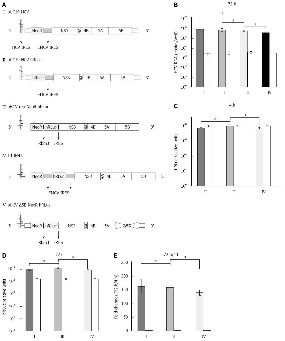
Figure 1 Replication and transgene expression of hepatitis C virus replicons.
A: Structure of hepatitis C virus (HCV) replicons. I: The parental replicon, pUC19-HCV, was a subgenomic replicon in which core, E1, E2, p7 and NS2 were replaced by neomycin phosphotransferase (NeoR) gene and encephalomyocarditis virus (EMCV) internal ribosome entry site (IRES); II: The bicistronic replicon, pUC19-HCV-hRLuc, was derived from pUC19-HCV by replacing NeoR with humanized Renilla luciferase (hRLuc) gene; III: The novel tricistronic replicon, pHCV-rep-NeoR-hRLuc, was derived from pUC19-HCV by replacing EMCV IRES with two sequential Rbm3 IRES and the hRLuc gene for simultaneous expression of NeoR and hRLuc.; IV: The tricistronic replicon with double EMCV IRESes, Tri-JFH1, was previously reported; V: The replication defective vector, pHCV-Δ5B-NeoR- hRLuc, was generated by truncating the active site of NS5B; B: Replication of HCV RNA in Huh-7 cells transiently transfected by replicons. Huh-7 cells were co-transfected with in vitro transcribed replicon RNA (I-IV) and replication-defective RNA from pHCV-Δ5B-NeoR-hRLuc as control (white columns). The replication of tricistronic HCV replicon was comparable to that derived from pUC19-HCV and pUC19-HCV-hRLuc and higher than that derived from Tri-JFH1; C: Transgene expression 4 h after replicon RNA transfection. The transgene of hRLuc was expressed at a significantly higher level in cells transfected with Rbm3 IRES tricistronic replicon than in cells transfected with pUC19-HCV-hRLuc and Tri-JFH1; D: Transgene expression 72 h after replicon RNA transfection; E: Fold changes of hRLuc activity between the time points of 4 and 72 h after transfection. aP < 0.05 between two groups.
- Citation: Cheng X, Gao XC, Wang JP, Yang XY, Wang Y, Li BS, Kang FB, Li HJ, Nan YM, Sun DX. Tricistronic hepatitis C virus subgenomic replicon expressing double transgenes. World J Gastroenterol 2014; 20(48): 18284-18295
- URL: https://www.wjgnet.com/1007-9327/full/v20/i48/18284.htm
- DOI: https://dx.doi.org/10.3748/wjg.v20.i48.18284


