Copyright
©2014 Baishideng Publishing Group Inc.
World J Gastroenterol. Dec 28, 2014; 20(48): 18228-18239
Published online Dec 28, 2014. doi: 10.3748/wjg.v20.i48.18228
Published online Dec 28, 2014. doi: 10.3748/wjg.v20.i48.18228
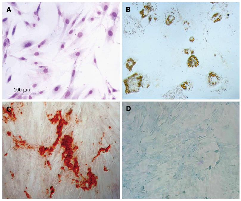
Figure 1 Mesenchymal stem cell characterization.
A: Mesenchymal stem cell (MSC) morphology by hematoxylin-eosin (HE) staining; B: Adipogenic differentiation detected by Oil red, which stains lipid vacuoles; C: Osteogenic differentiation detected by Alizarin Red, which stains deposit of calcium; D: Chondrogenic differentiation, proteoglycans stained by Alcian blue.
-
Citation: Gonçalves FDC, Schneider N, Pinto FO, Meyer FS, Visioli F, Pfaffenseller B, Lopez PLDC, Passos EP, Cirne-Lima EO, Meurer L, Paz AH. Intravenous
vs intraperitoneal mesenchymal stem cells administration: What is the best route for treating experimental colitis? World J Gastroenterol 2014; 20(48): 18228-18239 - URL: https://www.wjgnet.com/1007-9327/full/v20/i48/18228.htm
- DOI: https://dx.doi.org/10.3748/wjg.v20.i48.18228


