Copyright
©2014 Baishideng Publishing Group Inc.
World J Gastroenterol. Dec 7, 2014; 20(45): 17254-17259
Published online Dec 7, 2014. doi: 10.3748/wjg.v20.i45.17254
Published online Dec 7, 2014. doi: 10.3748/wjg.v20.i45.17254
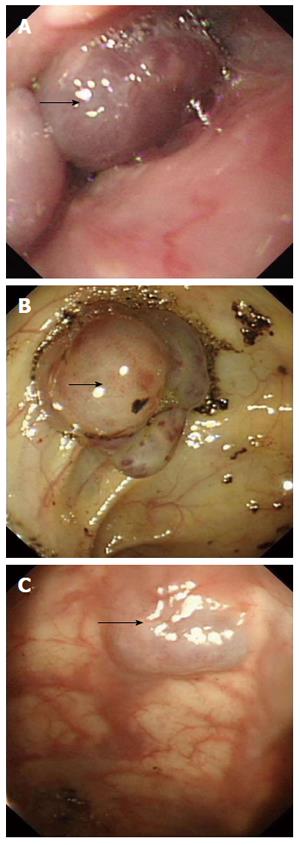
Figure 2 On endoscopy, glottis and esophagus (A) showed multiple bluish hemangiomatas (arrow), and no bleeding was seen; one lesion (2.
0 cm × 2.5 cm) with no fresh bleeding was seen in the colon (B) (arrow), and lesions were also observed on anus (C) (arrow).
- Citation: Jin XL, Wang ZH, Xiao XB, Huang LS, Zhao XY. Blue rubber bleb nevus syndrome: A case report and literature review. World J Gastroenterol 2014; 20(45): 17254-17259
- URL: https://www.wjgnet.com/1007-9327/full/v20/i45/17254.htm
- DOI: https://dx.doi.org/10.3748/wjg.v20.i45.17254


