Copyright
©2014 Baishideng Publishing Group Inc.
World J Gastroenterol. Dec 7, 2014; 20(45): 16964-16975
Published online Dec 7, 2014. doi: 10.3748/wjg.v20.i45.16964
Published online Dec 7, 2014. doi: 10.3748/wjg.v20.i45.16964
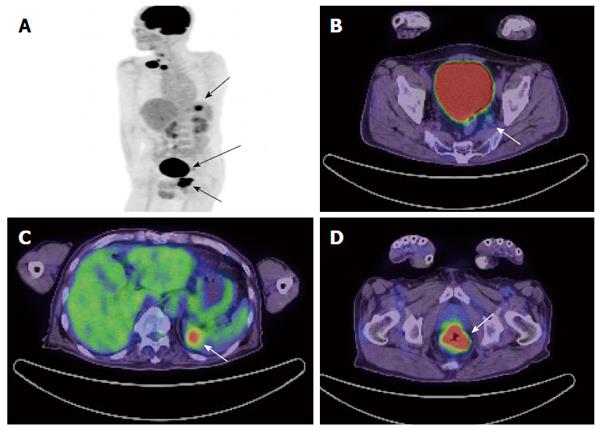
Figure 10 Sixty-four-year-old male with metastatic rectal cancer.
A: Maximum intensity projection image of PET shows the primary rectal lesion, metastasis of lateral lymph node and metastasis of spleen (arrow); B: PET/CT axial image shows a pathologically-confirmed metastasis of lateral lymph node with a SUVmax = 2.2 and diameter of 8 mm (arrow); C: PET/CT axial image shows metastasis of spleen (arrow); D: PET/CT axial image show primary rectal lesion (arrow). PET: Positron emission tomography; CT: Computed tomography; SUV: Standardized uptake value.
- Citation: Kijima S, Sasaki T, Nagata K, Utano K, Lefor AT, Sugimoto H. Preoperative evaluation of colorectal cancer using CT colonography, MRI, and PET/CT. World J Gastroenterol 2014; 20(45): 16964-16975
- URL: https://www.wjgnet.com/1007-9327/full/v20/i45/16964.htm
- DOI: https://dx.doi.org/10.3748/wjg.v20.i45.16964


