Copyright
©2014 Baishideng Publishing Group Inc.
World J Gastroenterol. Dec 7, 2014; 20(45): 16935-16947
Published online Dec 7, 2014. doi: 10.3748/wjg.v20.i45.16935
Published online Dec 7, 2014. doi: 10.3748/wjg.v20.i45.16935
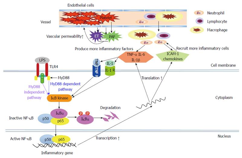
Figure 1 Schematic presentation of the pathways involved in the inflammatory response during acute pancreatitis.
Following lipopolysaccharide (LPS) stimulation, the inhibitors of κB (IκB) kinase are activated via the toll-like receptor 4 (TLR4)-myeloid differentiation factor 88 (MyD88)-dependent (blue) or -independent pathway (purple). Thereafter, IκBα is rapidly phosphorylated by IκB kinase and then degraded. This process allows nuclear factor-κB (NF-κB) to translocate into the nucleus and to increase the transcription of several important inflammatory genes, such as the genes encoding tumor necrosis factor-α (TNF-α), interleukin-1 (IL-1), adhesion molecules and chemokines. The up-regulation of these inflammatory mediators, in turn, either leads to further IκB kinase activation or mediates the migration of the inflammatory cells to the site of inflammation, thus amplifying the inflammatory response. Meanwhile, platelet-activating factor (released from active macrophages, endothelial cells or platelets) and endothelins (released from endothelial cells) are involved in the increased vascular permeability and extravasation of inflammatory cells. TNF R: Tumor necrosis factor receptor; IL-1 R: Interleukin-1 receptor; ICAM-1: Intercellularadhesionmolecule-1; PAF: Platelet-activating factor; ET: Endothelin.
- Citation: Li J, Yang WJ, Huang LM, Tang CW. Immunomodulatory therapies for acute pancreatitis. World J Gastroenterol 2014; 20(45): 16935-16947
- URL: https://www.wjgnet.com/1007-9327/full/v20/i45/16935.htm
- DOI: https://dx.doi.org/10.3748/wjg.v20.i45.16935


