Copyright
©2014 Baishideng Publishing Group Inc.
World J Gastroenterol. Nov 28, 2014; 20(44): 16786-16792
Published online Nov 28, 2014. doi: 10.3748/wjg.v20.i44.16786
Published online Nov 28, 2014. doi: 10.3748/wjg.v20.i44.16786
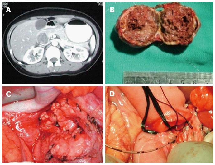
Figure 1 B-ultrasonic examination and computed tomography scan of case 1.
A: Enhanced computed tomography image of BTPH shows a low density in the pancreatic head mass at 3.5 cm × 3.2 cm. The tumor displayed ill-defined borderline in calcified lesions; B: Shape and structure of BTPH. The tumor is an oval shape and some necrotic tissue existed inside the tumor; C: Wound surface area of the pancreas. Measuring wound surface area of the pancreas is 6.3 cm × 6.5 cm after the tumor removed; D: Illustration for operative repair with Roux-en-Y pancreaticojejunostomy is shown with proximal pancreatic end ligated and a stent is put into distal pancreatic resection. BTPH: Benign tumors of the pancreatic head.
- Citation: Yuan CH, Tao M, Jia YM, Xiong JW, Zhang TL, Xiu DR. Duodenum-preserving resection and Roux-en-Y pancreatic jejunostomy in benign pancreatic head tumors. World J Gastroenterol 2014; 20(44): 16786-16792
- URL: https://www.wjgnet.com/1007-9327/full/v20/i44/16786.htm
- DOI: https://dx.doi.org/10.3748/wjg.v20.i44.16786


