Copyright
©2014 Baishideng Publishing Group Inc.
World J Gastroenterol. Nov 28, 2014; 20(44): 16690-16697
Published online Nov 28, 2014. doi: 10.3748/wjg.v20.i44.16690
Published online Nov 28, 2014. doi: 10.3748/wjg.v20.i44.16690
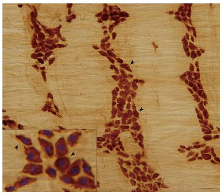
Figure 2 Representative light micrographs of whole mount preparations of the rat descending colon after HuC/HuD immunohistochemistry.
Arrowheads point to stained myenteric neurons. Density of neurons per field was determined with Plexus Pattern Analysis software, where the soma of the neurons was encircled (insert). Bars: 50 μm and 25 μm (insert).
- Citation: Talapka P, Nagy LI, Pál A, Poles MZ, Berkó A, Bagyánszki M, Puskás LG, Fekete &, Bódi N. Alleviated mucosal and neuronal damage in a rat model of Crohn’s disease. World J Gastroenterol 2014; 20(44): 16690-16697
- URL: https://www.wjgnet.com/1007-9327/full/v20/i44/16690.htm
- DOI: https://dx.doi.org/10.3748/wjg.v20.i44.16690


