Copyright
©2014 Baishideng Publishing Group Inc.
World J Gastroenterol. Nov 21, 2014; 20(43): 16368-16371
Published online Nov 21, 2014. doi: 10.3748/wjg.v20.i43.16368
Published online Nov 21, 2014. doi: 10.3748/wjg.v20.i43.16368
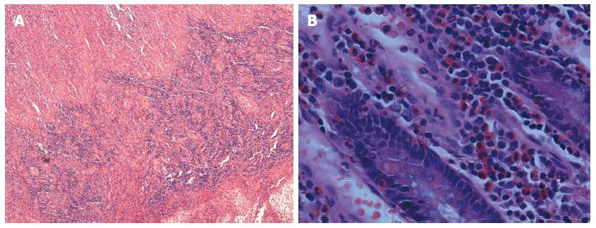
Figure 3 Microscopy of the resected ileocecal revealing eosinophilic infiltration from the submucosa to the subserosa.
A: Boundary of muscular and serosal layer become indistinct (HE × 40); B: Dense eosinophilic infiltrates in the mucosa (HE, × 400).
- Citation: Dai YX, Shi CB, Cui BT, Wang M, Ji GZ, Zhang FM. Fecal microbiota transplantation and prednisone for severe eosinophilic gastroenteritis. World J Gastroenterol 2014; 20(43): 16368-16371
- URL: https://www.wjgnet.com/1007-9327/full/v20/i43/16368.htm
- DOI: https://dx.doi.org/10.3748/wjg.v20.i43.16368


