Copyright
©2014 Baishideng Publishing Group Inc.
World J Gastroenterol. Nov 21, 2014; 20(43): 16203-16214
Published online Nov 21, 2014. doi: 10.3748/wjg.v20.i43.16203
Published online Nov 21, 2014. doi: 10.3748/wjg.v20.i43.16203
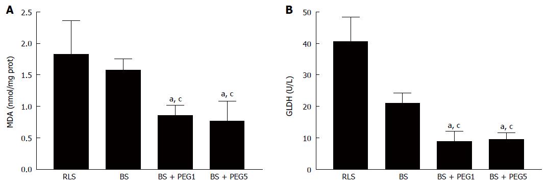
Figure 3 Hepatic malondialdehyde (A) and glutamate dehydrogenase (B) in livers rinsed with different washout solutions (RLS, BS, BS + PEG1, and BS + PEG5) and subjected to 2 h of normothermic reperfusion.
aP < 0.05 vs RLS; cP < 0.05 vs BS. PEG: Polyethylene glycol; RLS: Ringer’s lactate solution; BS: Base solution; BS + PEG1: BS with PEG-35 at 1 g/L; BS + PEG5: BS with PEG-35 at 5 g/L. MDA: Malondialdehyde; GLDH: Glutamate dehydrogenase.
- Citation: Zaouali MA, Bejaoui M, Calvo M, Folch-Puy E, Pantazi E, Pasut G, Rimola A, Ben Abdennebi H, Adam R, Roselló-Catafau J. Polyethylene glycol rinse solution: An effective way to prevent ischemia-reperfusion injury. World J Gastroenterol 2014; 20(43): 16203-16214
- URL: https://www.wjgnet.com/1007-9327/full/v20/i43/16203.htm
- DOI: https://dx.doi.org/10.3748/wjg.v20.i43.16203


