Copyright
©2014 Baishideng Publishing Group Inc.
World J Gastroenterol. Nov 14, 2014; 20(42): 15931-15936
Published online Nov 14, 2014. doi: 10.3748/wjg.v20.i42.15931
Published online Nov 14, 2014. doi: 10.3748/wjg.v20.i42.15931
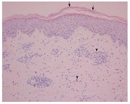
Figure 2 Hematoxylin and eosin stained erupted skin biopsy sample.
The epidermis is acanthotic with focal parakeratosis (arrows). A perivascular lymphocytic infiltrate admixed with interstitial eosinophils is present in the papillary and reticular dermis (arrowheads). Magnification: 200 ×.
- Citation: Kim JT, Jeong HW, Choi KH, Yoon TY, Sung N, Choi YK, Kim EH, Chae HB. Delayed hypersensitivity reaction resulting in maculopapular-type eruption due to entecavir in the treatment of chronic hepatitis B. World J Gastroenterol 2014; 20(42): 15931-15936
- URL: https://www.wjgnet.com/1007-9327/full/v20/i42/15931.htm
- DOI: https://dx.doi.org/10.3748/wjg.v20.i42.15931


