Copyright
©2014 Baishideng Publishing Group Inc.
World J Gastroenterol. Nov 14, 2014; 20(42): 15910-15915
Published online Nov 14, 2014. doi: 10.3748/wjg.v20.i42.15910
Published online Nov 14, 2014. doi: 10.3748/wjg.v20.i42.15910
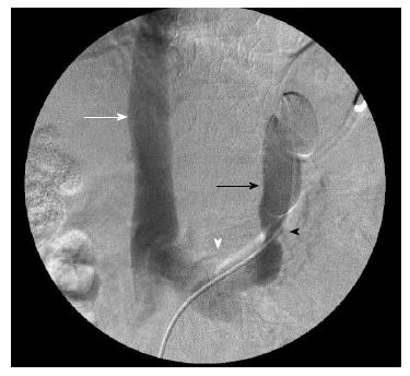
Figure 1 Venographic images of the splenorenal shunt before the embolization, showing a narrowing point where the 8 microcoils would be placed.
Inferior vena cava (white arrow); left renal vein (white arrowhead); splenorenal shunt with the catheter inside it (black arrow); narrowing location inside the splenorenal shunt (black arrowhead).
- Citation: Franzoni LC, Carvalho FC, Garzon RGA, Yamashiro FDS, Augusti L, Santos LAA, Dorna MS, Baima JP, Lima TB, Caramori CA, Silva GF, Romeiro FG. Embolization of splenorenal shunt associated to portal vein thrombosis and hepatic encephalopathy. World J Gastroenterol 2014; 20(42): 15910-15915
- URL: https://www.wjgnet.com/1007-9327/full/v20/i42/15910.htm
- DOI: https://dx.doi.org/10.3748/wjg.v20.i42.15910


