Copyright
©2014 Baishideng Publishing Group Inc.
World J Gastroenterol. Oct 28, 2014; 20(40): 14760-14777
Published online Oct 28, 2014. doi: 10.3748/wjg.v20.i40.14760
Published online Oct 28, 2014. doi: 10.3748/wjg.v20.i40.14760
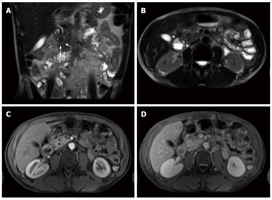
Figure 21 Segmental form groove pancreatitis.
Coronal (A) and axial (B) single-shot turbo spin-echo T2-weighted (HASTE) images with fat-suppression. Axial post-Gadolinium 3D-GRE T1-weighted images with fat-suppression during the hepatic arterial-dominant (C) and interstitial phases (D). There is mild duodenal thickening and edema (arrowheads) (A, B) associated with a sheet-like mass in the pancreaticoduodenal groove that extends to the pancreatic head and demonstrates decreased T1 signal pre-contrast (not shown), slightly decreased T2 signal with tiny cystic changes (white arrows) (A, B), early arterial hypo-enhancement (black arrow) (C) and progressive enhancement on the subsequent delayed images (D) due to fibrosis, in keeping with segmental form groove pancreatitis.
- Citation: Manikkavasakar S, AlObaidy M, Busireddy KK, Ramalho M, Nilmini V, Alagiyawanna M, Semelka RC. Magnetic resonance imaging of pancreatitis: An update. World J Gastroenterol 2014; 20(40): 14760-14777
- URL: https://www.wjgnet.com/1007-9327/full/v20/i40/14760.htm
- DOI: https://dx.doi.org/10.3748/wjg.v20.i40.14760


