
















©2014 Baishideng Publishing Group Inc.
World J Gastroenterol. Oct 28, 2014; 20(40): 14760-14777
Published online Oct 28, 2014. doi: 10.3748/wjg.v20.i40.14760
Published online Oct 28, 2014. doi: 10.3748/wjg.v20.i40.14760
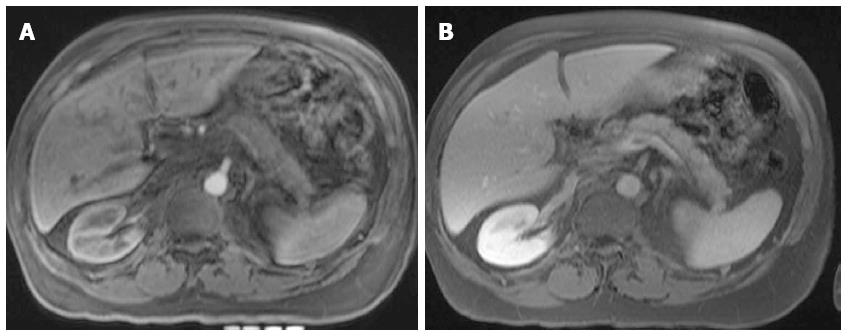
Figure 15 Chronic pancreatitis.
A: Axial post-Gadolinium 3D-GRE T1-weighted image with fat-suppression during the hepatic arterial-dominant phase; B: Axial post-Gadolinium FLASH in-phase T1-weighted image with fat-suppression during the hepatic-venous phases. The pancreatic body and tail show poor early contrast enhancement (A) and mild enhancement on the delayed images related to fibrosis (B) in keeping with chronic pancreatitis.
- Citation: Manikkavasakar S, AlObaidy M, Busireddy KK, Ramalho M, Nilmini V, Alagiyawanna M, Semelka RC. Magnetic resonance imaging of pancreatitis: An update. World J Gastroenterol 2014; 20(40): 14760-14777
- URL: https://www.wjgnet.com/1007-9327/full/v20/i40/14760.htm
- DOI: https://dx.doi.org/10.3748/wjg.v20.i40.14760