
















©2014 Baishideng Publishing Group Inc.
World J Gastroenterol. Oct 21, 2014; 20(39): 14500-14504
Published online Oct 21, 2014. doi: 10.3748/wjg.v20.i39.14500
Published online Oct 21, 2014. doi: 10.3748/wjg.v20.i39.14500
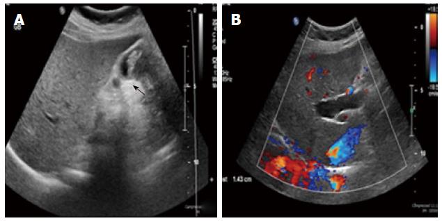
Figure 1 Ultrasound.
A: Ultrasound showing thick-walled gallbladder and intraluminal mass (arrow); B: Ultrasound showing right intrahepatic bile duct dilation.
- Citation: Rungsakulkij N, Boonsakan P. Synchronous gallbladder and pancreatic cancer associated with pancreaticobiliary maljunction. World J Gastroenterol 2014; 20(39): 14500-14504
- URL: https://www.wjgnet.com/1007-9327/full/v20/i39/14500.htm
- DOI: https://dx.doi.org/10.3748/wjg.v20.i39.14500