Copyright
©2014 Baishideng Publishing Group Inc.
World J Gastroenterol. Oct 14, 2014; 20(38): 14063-14067
Published online Oct 14, 2014. doi: 10.3748/wjg.v20.i38.14063
Published online Oct 14, 2014. doi: 10.3748/wjg.v20.i38.14063
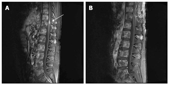
Figure 1 T1-weighted magnetic resonance image of the spine.
A: The 5.9 mm × 5.3 mm × 14.8 mm metastatic tumor was observed in the T10/11 level (arrow). Diffuse vertebral metastases were also observed in almost the entire spine; B: The intramedullary tumor was removed by surgical treatment.
- Citation: Kim JH, Hyun CL, Han SH. Intramedullary spinal cord metastasis from pancreatic neuroendocrine tumor. World J Gastroenterol 2014; 20(38): 14063-14067
- URL: https://www.wjgnet.com/1007-9327/full/v20/i38/14063.htm
- DOI: https://dx.doi.org/10.3748/wjg.v20.i38.14063


